Sickle Cell Anemia Investigation: Day 4
Today began with a video by John Perry’s series “Stated Clearly” titled “What is a gene?”
Next, students wrote down the learning objective. A protein’s shape determines its function, and the shape occurs due to the sequence of amino acids coded by DNA. I explained that a truck has a specific shape that helps it to do its function of hauling things and a ferrari has a different shape that helps it to do its function of going fast. Proteins too have specific shapes to help them do their own function.
It is a difficult abstraction for students to take to look at a protein model and think that these microscopic things actually have a function.

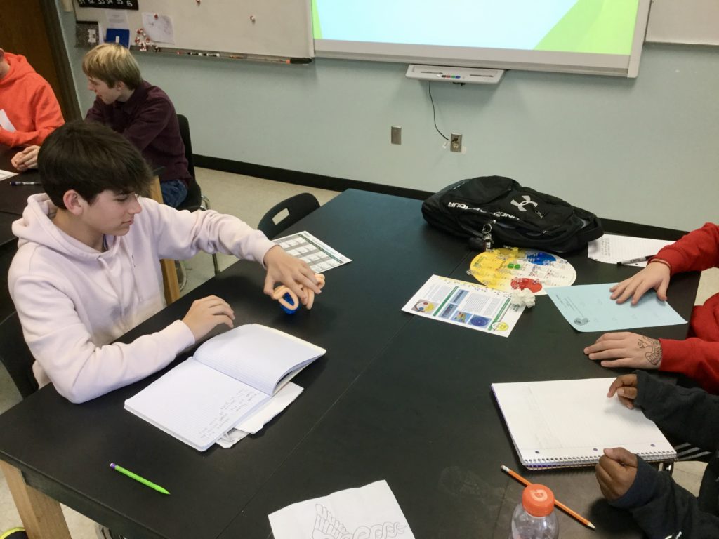
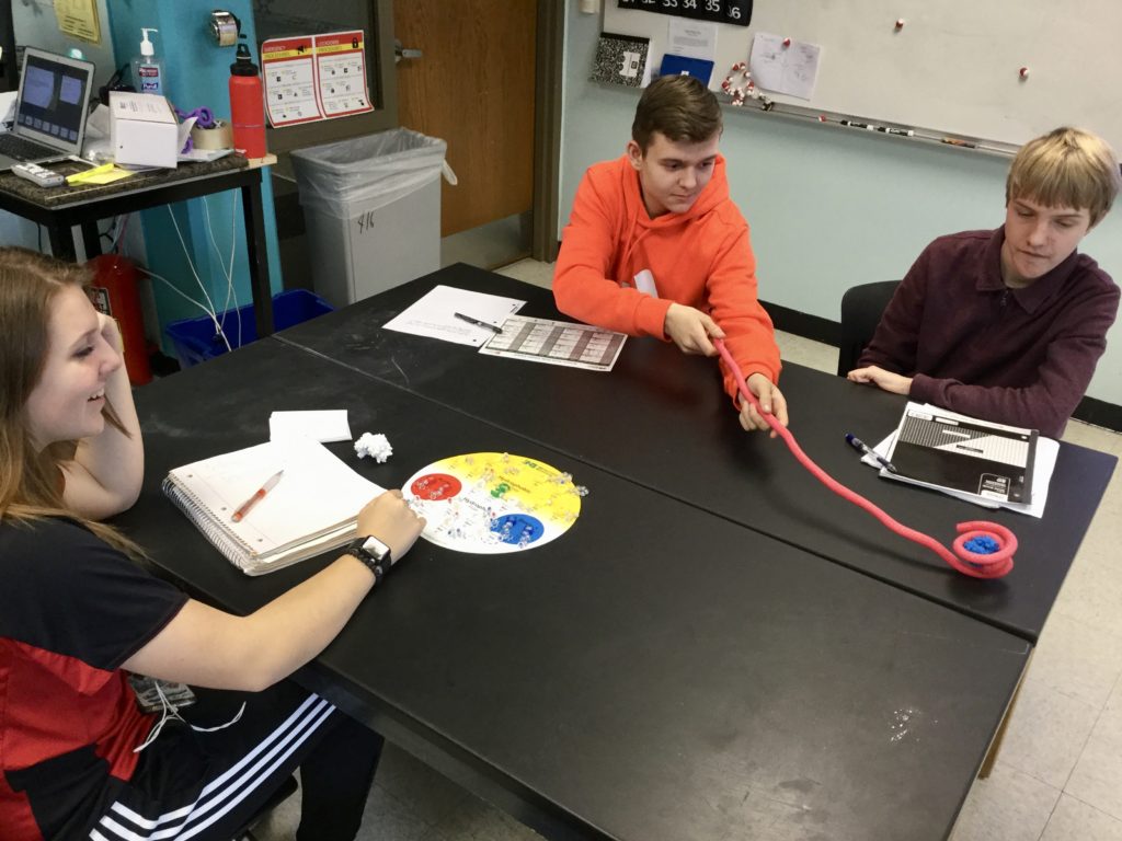
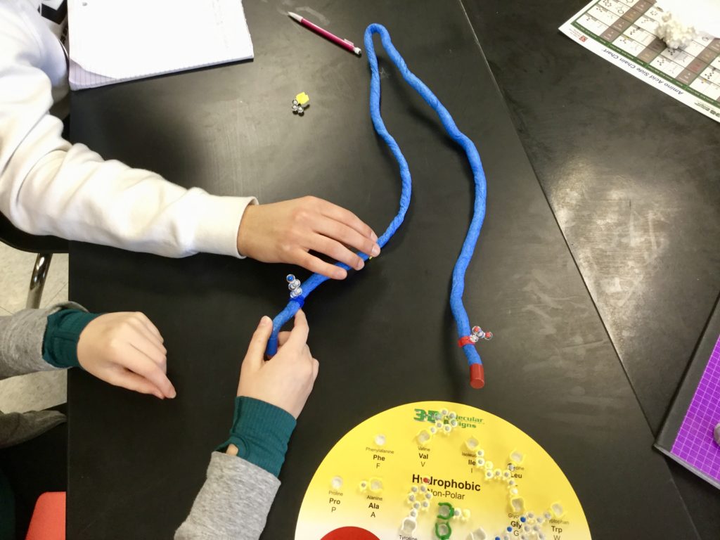
In order to help students realize how these proteins function can work I use minitoobers. (*this is not my own original idea.) I tell students that they have to fold their protein using beta pleated sheets a.k.a. zigzags or alpha helicies a.k.a. loops into a shape that can pick up a molecule and transfer it from one partner to the next. I give them 3 minutes to fold their toober into a shape that can make the transfer and then let them show the rest of the class their solution.
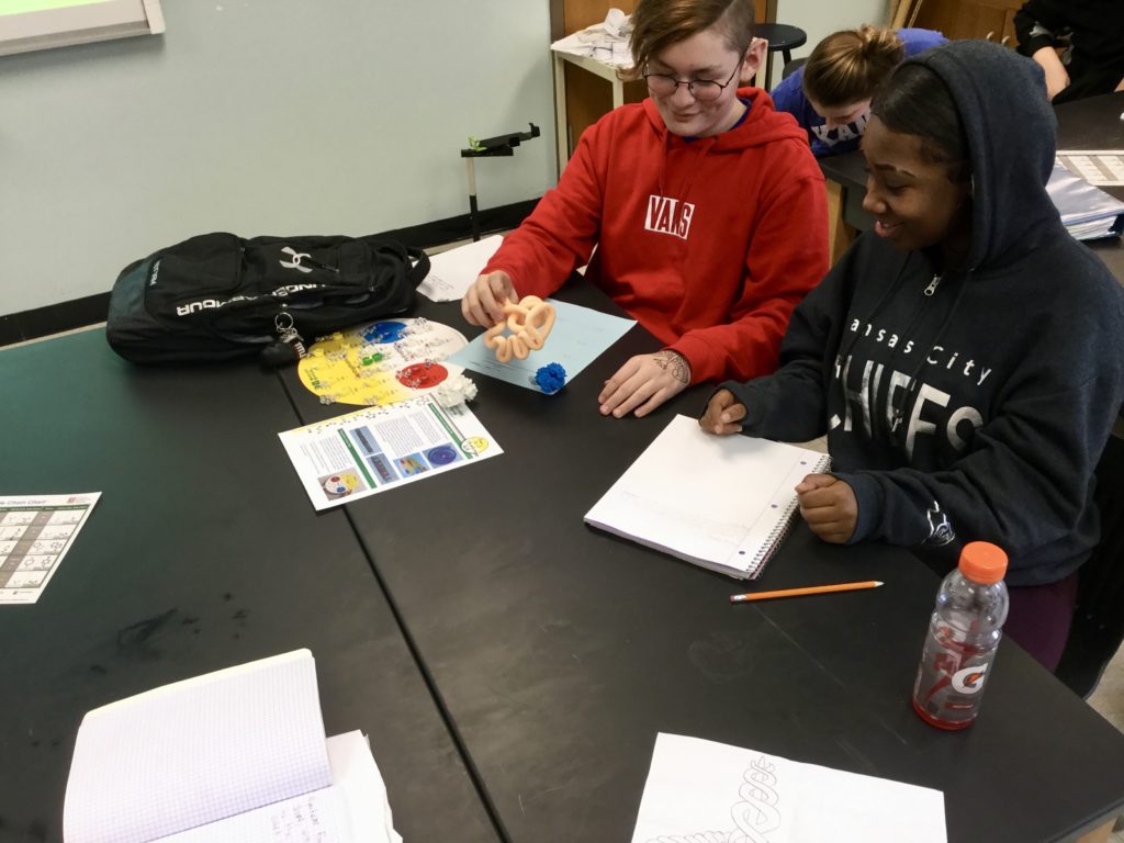
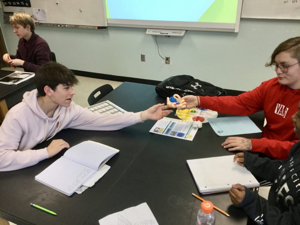
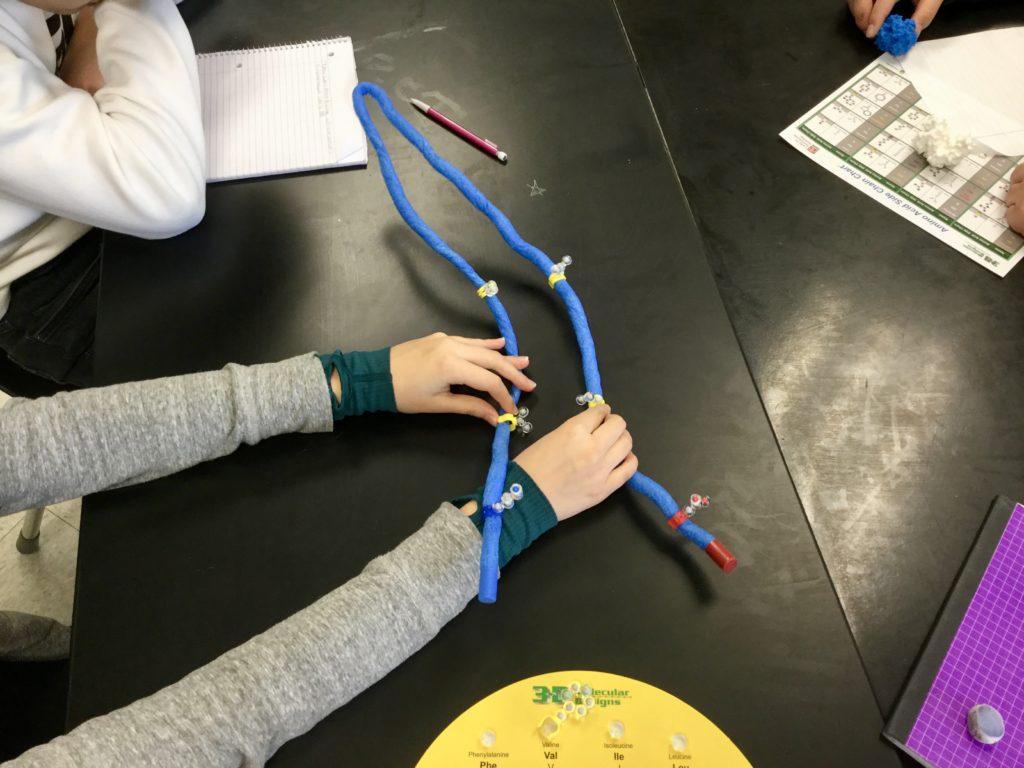
Students fold their proteins into many different shapes to perform the function of moving a protein.


I can then show them It turns out these protein things can have many shapes! You can show the kids these proteins too from the Protein Data Bank.
Now we turn our attention to how the sequence of DNA effects the shape of a protein. I am using kits that we purchased from 3dmolecular designs. But, colored push pins will work too. ( I think Drew Ising did a post about this in the past).
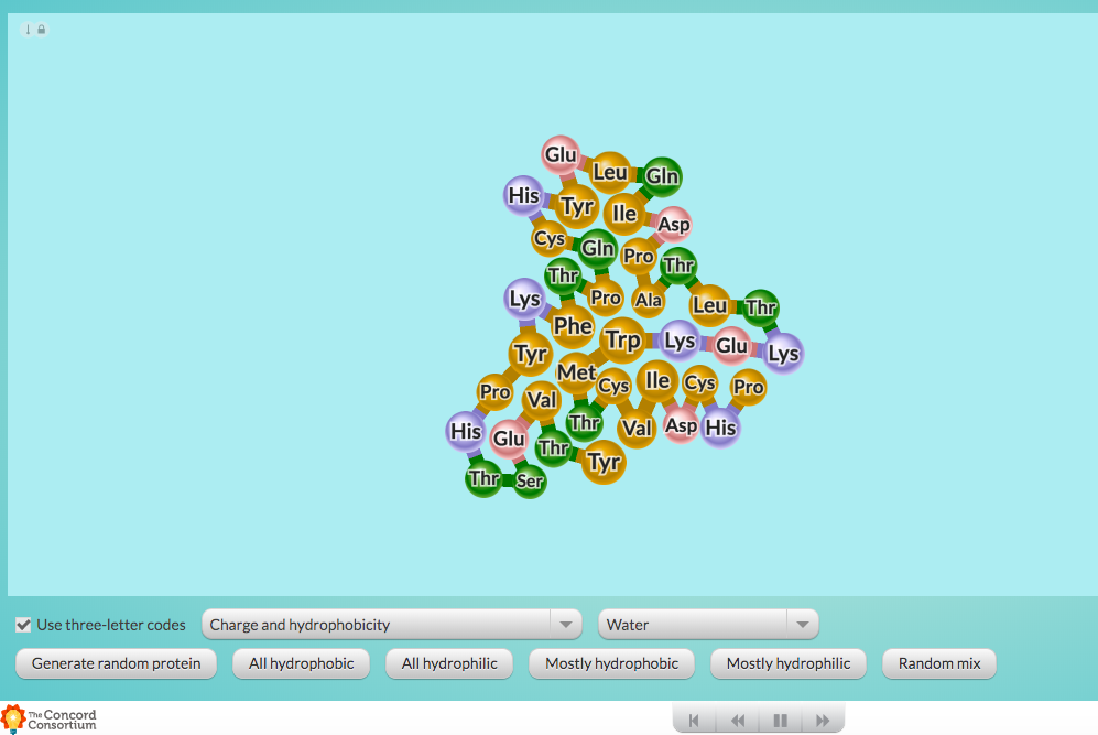
At this point I pause the class and open a concord consortium interactive player on my smartboard.

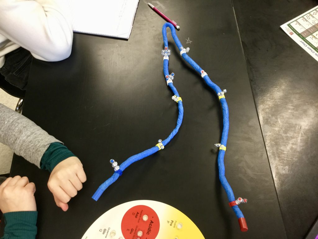
Now it is the students chance to fold the protein into a shape where the positive and negative amino acids on the ends of their toober attract to one another and the hydrophobic amino acids fold in.
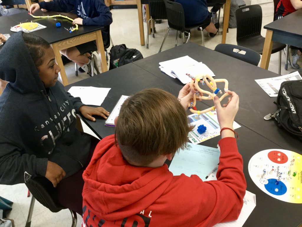
At this point I grab one of the students folded protein and I substitute one positive amino acid for a negative amino acid. They can see that it will unravel their protein. Now I try to hit home the reason that a mutation in DNA can cause an impact in an organism.
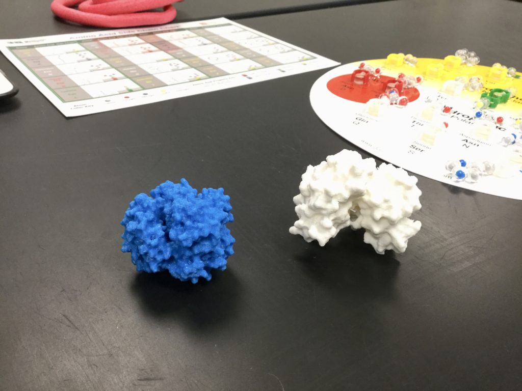
I had a few Hemoglobin molecules printed off for my classroom. I found the STL files on Thingverse. So, now the kids can see the shape of the protein is different. When only one amino acid is changed.
 Previous Post
 Next Post