Day 1: Fly Genetics
In an effort to channel Brad earlier this fall, I will (hopefully) be blogging about an ongoing interdisciplinary project a researcher and I are currently working on. The blog posts are hosted also on my blog and therefore may contain odd wording or obvious/redundant information, but partly this is for my memory as well as an audience possibly unfamiliar with genetics. All photos are mine or credited to their owner and any information published is with the permission of my researcher as well as we hope to publish our approach as a way to illustrate collaboration between secondary and post-secondary institutions. Enjoy:
Today marks the beginning of what I think it going to be a really cool project for my students. Through a connection at at the University of Kansas I and a PhD candidate my students (both freshman and AP biology students) will be embarking on an 2-month interdisciplinary project.
Basically, students will be attempting to characterize a protein involved in epithelial cell stabilization (specifically a protein in the septate junction of Drosophila melanogaster).
Today was technically day 1 (the researcher came by last Monday and met the students and introduced their research focus and what they’ll be doing as an overview).
First, students tried to recall Drosophila epithelial cell structure and function, which surprisingly they all remembered, at least the essential components. They remembered how to draw the cells and that there were “walls” involved that separated the chemicals from the apical (outside) and basal (inside) regions. The names we can hammer in later, but that conceptual understanding was nice to see.
We then had students go to their dissecting scopes to observe their embryos for the first time. This was also just the second time students had used dissecting scopes this year so they were getting a lot of new experiences. Students were viewing multiple embryos on apple juice agar plates with yeast (for food).

apple juice plate w/ yeast paste on right, flies on left
Students were given the task of drawing what they saw (we did not tell them they were fly embryos, just to look and observe) using a petri dish outline in their composition notebooks for drawing their field of view. Of course, I showed students once again the awesomeness of their camera-phones which did it’s usual trick of instantly turning some students into microphotographers! Here is one of the better photos:

- 30x magnification
Can you see their dorsal appendages?

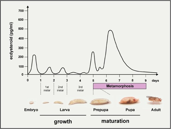
So after students decided we had embryos, we showed them the rest of the fly life cycle (below) and asked, what do you expect to see tomorrow? They decided they would see larva and we asked what they expected to larva to do? Once they said moving some students shouted, “Some of mine are moving!” Then we had that student draw what they saw of the moving fly and it got most others excited for some movement tomorrow. What was fun (I thought) was when we asked students based on this molt hormone’s levels through the flies life, what stage of human life correlates with the giant spike in hormones for pupa? (A: PUBERTY! Gotta love that reaction from 14 years olds)

- Drosophila Life Cycle (w/ a molt hormone levels throughout)
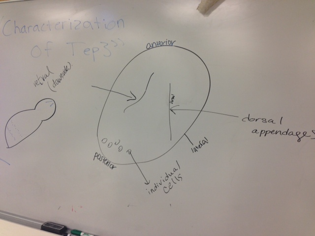
Then the last thing we did was we had volunteers one by one draw components (example below) of the Drosophila anatomy, so that we could use a common terminology when communicating throughout the project. For the freshman it may have involved mimicing antlers to remember anterior and even one dedicated teacher lying on a table dorsal side up (image not found) so students could visualize how they were viewing their embryos from their dissecting scopes.
- student fly anatomy drawing
So with that (rinse, repeat 5 times), day one was complete and day two is tomorrow! Stay tuned!

Nice post Camden! I will be following along…